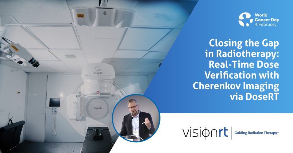
Closing the Gap in Radiotherapy: Real-Time Dose Verification with Cherenkov Imaging via DoseRT
World Cancer Day is a moment to reflect on how advances in cancer care translate into safer, more effective treatment for patients. In radiotherapy, this progress depends not only on how precisely treatments are planned, but on how confidently they can be verified as they are delivered.
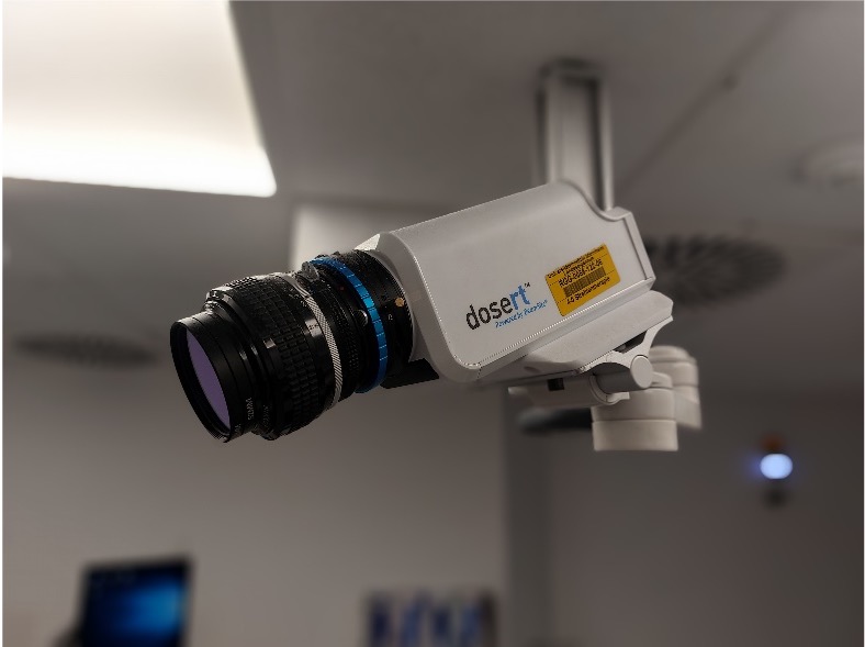
At the University Medical Center Mannheim, University of Heidelberg, Germany, this focus on verification during treatment was a key motivation for Dr. Florian Stieler and his team to adopt a new technology into their clinical practice. “We introduced DoseRT as a Cherenkov imaging device to address an information gap in radiotherapy,” he explains. “While image guidance tools provide information on patient positioning prior to treatment and patient motion during radiation therapy, and conventional QA tools assess machine performance in separate non-clinical sessions, these tools do not capture the dose delivery during actual patient treatment. Cherenkov imaging provides real-time visualization of radiation delivery, enabling detection of patient setup errors, patient motion and unintended irradiation“.
By bringing this missing information together during treatment, DoseRT provides insight that was previously unavailable. “All of this information comes together in one place,” says Dr. Stieler. “For the first time, we can see what we are actually delivering, while we deliver it.”

Clinical Experience with Cherenkov Imaging via DoseRT
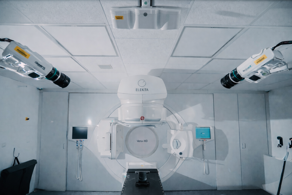
DoseRT was introduced without disrupting established clinical workflows. “Cherenkov imaging with DoseRT is integrated with the AlignRT system to monitor dose delivery and patient positioning in real-time,” says Dr Stieler. In daily practice, this visibility has proven clinically valuable. “DoseRT has been valuable to identify deviations in radiotherapy between planned and delivered dose caused by changes in patient anatomy, position and motion, and linac performance,” he explains. This information, which previously existed only as indirect or surrogate data, is now visible during treatment itself.
“Breast treatments, especially with tangential beams, are suited for Cherenkov imaging because of the proximity of the target to the patient’s surface and the resulting intuitive Cherenkov intensity map,” he says. The signal readily reveals inhomogeneous coverage or unintended irradiation of the contralateral breast or axillary region.
However, at his clinic, the focus extends well beyond breast treatments. “We analyze all indications,” he explains, “especially complex and high-dose paradigms like SBRT, head and neck irradiation, or stereotactic arrhythmia radioablation.” These complex cases, also presented at the 2025 European SGRT Community Meeting, are where confidence in delivery is most critical.
The Future of Real-Time Dose Verification in Radiotherapy
Looking ahead, Dr Stieler sees Cherenkov imaging becoming a standard part of everyday radiotherapy practice. “The future of Cherenkov imaging lies in its routine integration into clinical workflows as a real-time verification and risk management tool that complements image guidance and quality assurance by providing direct feedback on treatment accuracy and delivered dose,” he says.
For clinics seeking greater transparency, confidence, and safety in radiotherapy delivery, DoseRT offers something that has long been missing: the ability to truly see what is being treated, as it happens.
Get in touch
Ready to take the next step?
Vision RT’s family of SGRT solutions guide radiation therapy for better patient care at every step: Sim, Planning, Treatment and Dose. Whether you’re looking for a quote, a product demo (virtual or in-person) or just more information, please get in touch.