Making the business case for SGRT with AlignRT

Making the business case for SGRT with AlignRT

“A wise investment” – Advisory Board

Radiation oncology centers are facing a number of challenges in today’s financial environment. Due to dynamic healthcare reimbursement and recent market changes, administrators are under ever-increasing pressure to ensure their investments will bring about better patient outcomes while minimizing total cost of care.

Efficiency, patient satisfaction, and competitive differentiation are key.

A recent oncology-focused technology brief from Advisory Board, a health care think tank with more than 4,500 member organizations, recommended use of Surface Guided Radiation Therapy (SGRT) as a “wise investment” for cancer centers. “Using SGRT,” the report stated, “as opposed to marker-based techniques, allows for improved accuracy, patient satisfaction, and throughput.”

Financial Health in a Value-based Healthcare Environment

15 of the top 15 “Best Hospitals for Cancer”, have Vision RT systems, according to the latest analysis by U.S. News and World Report. And as centers increasingly move to hypofractionated treatments, the ability to monitor patients in real-time is more important than ever. Motion management through SGRT is becoming an essential safety element in patient treatment delivery across the globe.

Vision RT is the market leader in SGRT. It’s a technology that we invented, and our system offers multiple advantages in a value-based reimbursement environment:

Improve your outcomes

Vision RT has published clinical evidence of eliminating cardiac perfusion defects after left breast cancer treatment; from 27% of patients to 0%2, 3

Reduce your cost of care

Published data demonstrating 14% time savings and streamlined workflows36  ; AlignRT system up-time: 98%

Enhance patient experience

AlignRT eliminates the need for closed masks and permanent marks or tattoos 36 ; reduces patient treatment times10 and the need for pediatrics anesthesia37 .

Mitigate your risks

It is estimated that approximately 50% of reported near misses / medical events38  occurring at the time of treatment could have been prevented using Vision RT’s technology.

Using Marketing Resources to Increase Patient Volume

Local marketing support

Recognizing the value of local patient marketing, Vision RT has developed +20 marketing assets (e.g., brochures, ads, scripts) for you to customize and use in your market to promote AlignRT/SGRT. Executing a local campaign can differentiate your center from your competition and drive patients to seek treatment at your center.

A number of these resources also highlight the tattoo and mark-free benefit of AlignRT use, if applicable at your clinic.

78 % of patients prefer a tattoo-less option 35


Case study – U.S. Midwestern Cancer Center

  • Using Vision RT marketing resources, one center executed a local marketing campaign that focused on the heart-sparing benefit of AlignRT for left-breast cancer patients. Assets used were website copy and images, a press release, print ads, social ads, and patient education brochures.
  • Measured results showed a 100% increase in patient volume (55 to 110) in less than 12 months.

Patients are willing to travel 45 additional miles for a center with a tattoo-less option 35
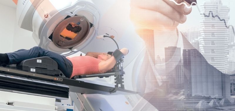
Vision RT can help you maximize your return on investment. With AlignRT adoption, centers have increased patient volume by as much as 20%


Data from a mid-west center using local marketing resources to increase patient volume

Radiation therapy technology that supports APM objectives

Efficiency gains with SGRT markerless positioning for breast radiotherapy

Hear from our users

  1. Heinzerling JH, et al. Use of surface-guided radiation therapy in combination with igrt for setup and intrafraction motion monitoring during stereotactic body radiation therapy treatments of the lung and abdomen. J Appl Clin Med Phys 2020.
  2. Marks et al. The incidence and functional consequences of RT‑associated cardiac perfusion defects. Int J Radiat Oncol Biol Phys. 2005 Sep 1;63(1):214-23.
  3. Zagar T, et al. Utility of Deep Inspiration Breath Hold for Left-Sided Breast Radiation Therapy in Preventing Early Cardiac Perfusion Defects: A Prospective Study. Int J Radiat Oncol Biol Phys 2017;97 (5):903-909.
  4. Gierga et al. A Voluntary Breath-Hold Treatment Technique for the Left Breast With Unfavorable Cardiac Anatomy Using Surface Imaging. Int J Radiat Oncol Biol Phys. 2012 Dec 1;84(5):e663-8
  5. Cerviño et al. Using surface imaging and visual coaching to improve the reproducibility and stability of deep-inspiration breath hold for left-breast-cancer radiotherapy. Phys Med Biol. 2009 Nov 21;54(22):6853-65.:
  6. Chang et al. Video surface image guidance for external beam partial breast irradiation. Pract Radiat Oncol. 2012 Apr-Jun;2(2):97-105.: :
  7. Padilla et al. Assessment of interfractional variation of the breast surface following conventional patient positioning for whole-breast radiotherapy. J Appl Clin Med Phys. 2014 Sep 8;15(5):4921:
  8. Rochet et al. Deep inspiration breath-hold technique in left sided breast cancer radiation therapy: Evaluating cardiac contact distance as a predictor of cardiac exposure for patient selection. Practical Radiation Oncology (2015) 5, e127-e134:
  9. Rong et al. Improving intra-fractional target position accuracy using a 3D surface surrogate for left breast irradiation using the respiratory-gated deep-inspiration breath-hold technique. PLoS One. 2014 May 22;9(5):e97933
  10. Shah et al. Clinical evaluation of interfractional variations for whole breast radiotherapy using 3-dimensional surface imaging. Pract Radiat Oncol. 2013 Jan-Mar;3(1):16-25.:
  11. Tang et al. Clinical experience with 3-dimensional surface matching-based deep inspiration breath hold for left-sided breast cancer radiation therapy. Pract Radiat Oncol. 2014 May-Jun;4(3):e151-8.:
  12. Tang et al. Dosimetric effect due to the motion during deep inspiration breath hold for left-sided breast cancer radiotherapy. J Appl Clin Med Phys. 2015 Jul 8;16(4):5358.:
  13. Tanguturi et al. Prospective assessment of deep inspiration breath-hold using 3-dimensional surface tracking for irradiation of left-sided breast cancer. Pract Radiat Oncol. 2015 Nov-Dec;5(6):358-65
  14. Pan H, et al. Frameless, real-time, surface imaging-guided radiosurgery: Clinical outcomes for brain metastases. Neurosurgery 2012;71 (4):844-851.
  15. Pham NL, et al. Frameless, real-time, surface imaging-guided radiosurgery: Update on clinical outcomes for brain metastases. Translational Cancer Research 2014;3 (4):351-357.
  16. Lau S et al. Clinical efficacy and safety of surface imaging guided radiosurgery (SIG-RS) in the treatment of benign skull base tumors. J Neurooncol (2017) 132:307–312
  17. Peng et al. Characterization of a real-time surface image-guided stereotactic positioning system. Med Phys. 2010 Oct;37(10):5421-33.
  18. Baker et al. Trigeminal Rhizotomy Performed with Modern Image-guided Linac: Case Report, Cureus, 5(9), 139, 2013
  19. Cerviño et al. Initial clinical experience with a frameless and maskless stereotactic radiosurgery treatment. Pract Radiat Oncol. 2012 Jan-Mar;2(1):54-62.
  20. Lau et al. Single-Isocenter Frameless Volumetric Modulated Arc Radiosurgery for Multiple Intracranial Metastases. Neurosurgery. 2015 Aug;77(2):233-40;
  21. Li et al. Clinical experience with two frameless stereotactic radiosurgery (fSRS) systems using optical surface imaging for motion monitoring. J Appl Clin Med Phys. 2015 Jul 8;16(4):5416.
  22. Li et al. Motion monitoring for cranial frameless stereotactic radiosurgery using video-based three-dimensional optical surface imaging. Med Phys. 2011 Jul;38(7):3981-94.
  23. Li et al. Optical Surface Imaging for Online Rotation Correction and Real‐Time Motion Monitoring with Threshold Gating for Frameless Cranial Stereotactic Radiosurgery, M9ed. Phys., Volume 38, 3711, 2011
  24. Mancosu et al. Accuracy evaluation of the optical surface monitoring system on EDGE linear accelerator in a phantom study. Med Dosim. 2016 Summer;41(2):173-9
  25. Pan et al. Frameless, real-time, surface imaging-guided radiosurgery: clinical outcomes for brain metastases. Neurosurgery. 2012 Oct;71(4):844-51.
  26. Pham et al. Frameless, real-time, surface imaging-guided radiosurgery: update on clinical outcomes for brain metastases. Trans. Cancer Res, 3, 4, 351-357, August, 2014.
  27. Wen et al. Characteristics of a novel treatment system for linear accelerator-based stereotactic radiosurgery. J Appl Clin Med Phys. 2015 Jul 8;16(4):5313.
  28. Wen et at. Technical Note: Evaluation of the systematic accuracy of a frameless, multiple image modality guided, linear accelerator based stereotactic radiosurgery system. Med Phys. 2016 May;43(5):2527,
  29. Wiersma et al. Spatial and temporal performance of 3D optical surface imaging for real-time head position tracking. Med Phys. 2013 Nov;40(11):111712.
  30. Cerviño et al. Frame-less and mask-less cranial stereotactic radiosurgery: a feasibility study. Phys Med Biol. 2010 Apr 7;55(7):1863-73
  31. Paravati et al. Initial clinical experience with surface image guided (SIG) radiosurgery for trigeminal neuralgia, Translational Cancer Research, 3, 4, 333-337, August, 2014,
  32. Baker et al. Trigeminal Rhizotomy Performed with Modern Image-guided Linac: Case Report, Cureus, 5(9), 139, 2013
  33. Wen N, et al. Technical note: Evaluation of the systematic accuracy of a frameless, multiple image modality guided, linear accelerator based stereotactic radiosurgery system. Med Phys 2016;43 (5):2527.
  34. Covington E, et al. Submillimeter monitoring of intrafraction patient movement with optical surface imaging. AAPM Annual Meeting 2018.
  35. The Breast Journal. Radiotherapy tattoos: Women’s skin as a carrier of personal memory—What do we cause by tattooing our patients? September 24, 2019 online publication. Accessed November 12, 2019 at https://onlinelibrary.wiley.com/doi/epdf/10.1111/tbj.13591
  36. Herron E, et al. Surface guided radiation therapy as a replacement for patient marks in treatment of breast cancer. Int J Radiat Oncol Biol Phys. 2018; 102 (3):e492-e493.
  37. Alisha Cheblick, B.S., R.T.(T), Childrens Hospital of Los Angeles. Presentation at SGRT Annual Meeting, Use of Surface Guidance in the Pediatric Setting. March 2017.
  38. From a global reporting database. Data analyzed by Vision RT from SAFRON database. https://www.iaea.org/resources/rpop/resources/databases-and-learning-systems/safron
  39. Flores-Martinez E, et al. Assessment of the use of different imaging and delivery techniques for cranial treatments on the halcyon linac. Journal of Applied Clinical Medical Physics 2020;21 (1):53-61

Get in touch

Ready to take the next step?

Vision RT’s family of SGRT solutions guide radiation therapy for better patient care at every step: Sim, Planning, Treatment and Dose. Whether you’re looking for a quote, a product demo (virtual or in-person) or just more information, please get in touch.